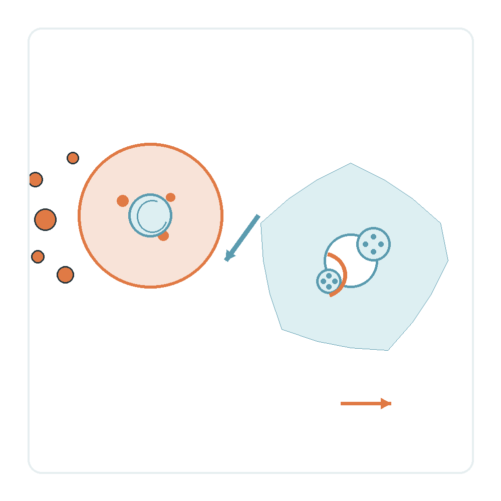
In brief: Imagine senescent cells in aging tissues as scrap parts waiting for pickup. When scrap accumulates, the bottleneck is rarely just an increased manufacturing rate. Often, the cleanup fleet itself has stalled. Published in Nature Aging in 2026, this study shows that the age-related decline of chaperone-mediated autophagy (CMA) simultaneously changes senescent cell secretions and paralyzes macrophage engulfment machinery.
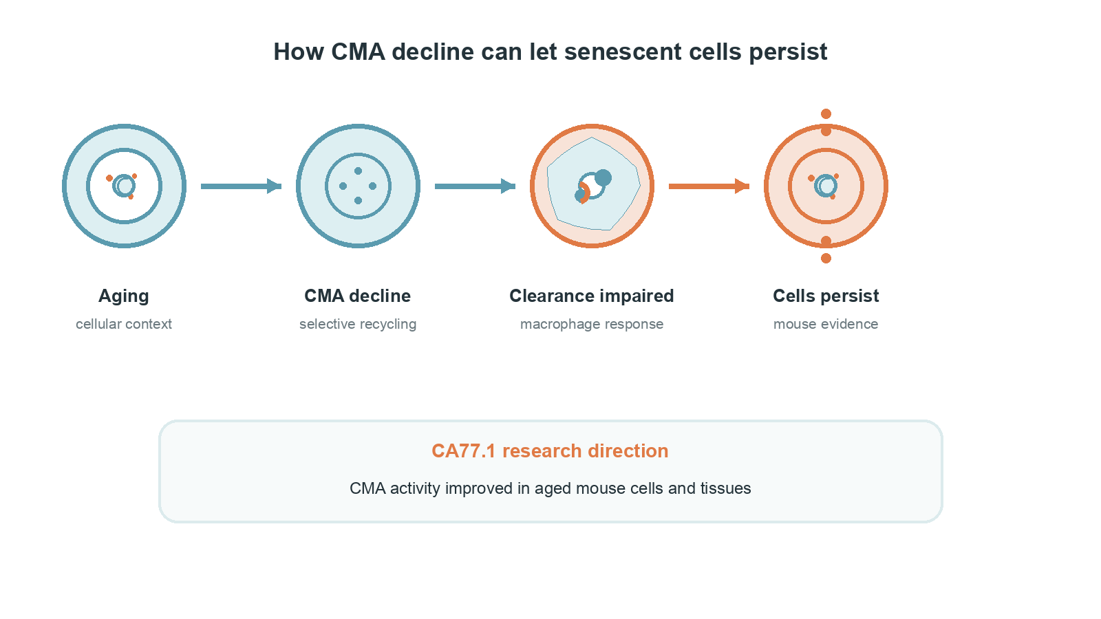
The short answer: aging stalls both ends of the clearance chain
Age-associated decline in CMA alters senescent-cell secretions and directly impairs macrophage engulfment capacity.
We often assume that senescent cells pile up in old tissues simply because more damaged cells undergo cell-cycle arrest. However, healthy macrophages are supposed to identify and swallow these cells promptly. The researchers compared mouse ear fibroblasts from 23-month-old and 4-month-old mice. Old fibroblasts showed significantly lower basal CMA activity. Moreover, when senescence was induced with palbociclib and evaluated after 7 days, young cells upregulated CMA, whereas aged cells completely failed to activate this recycling response.
Importantly, CMA deficiency alone did not transform healthy cells into fully senescent cells. Instead, it reshaped the proteomic, metabolic, and secretory profiles of senescent cells. The resulting secretome exhibited more aggressive pro-senescence characteristics and actively suppressed macrophage function, leaving tissue clearance stalled at both ends.
CMA is not a master switch, but a selective recycling line in the lysosome
CMA operates as a precision lysosomal degradation route guided by HSC70 substrate recognition and LAMP2A docking.
Unlike bulk macroautophagy that engulfs broad cytoplasm portions, CMA functions like an optical sorting line in a recycling workshop. The cytosolic chaperone HSC70 scans for proteins bearing a specific KFERQ-like motif. Upon recognition, HSC70 shuttles these targeted substrates directly to the lysosomal membrane receptor LAMP2A for translocation and degradation.
Because this pathway acts as a dedicated maintenance team for critical regulatory proteins, its age-related decline creates targeted proteomic imbalances. When damaged or inhibitory signaling proteins are not dismantled in time, cellular regulation loses its equilibrium.
Why macrophages see senescent cells but fail to engulf them
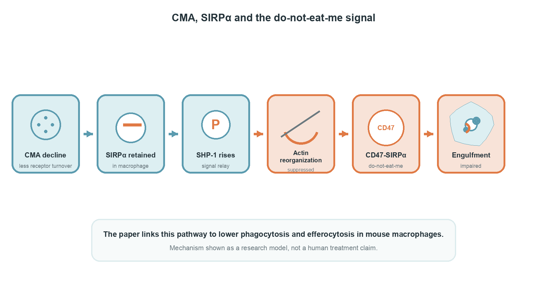
Macrophage CMA deficiency blocks SIRPα degradation, driving SHP-1 retention and suppressing the actin reorganization needed for phagocytosis.
Macrophages represent the frontline cleaners tasked with clearing dead and senescent cells. Why do they fail to engulf senescent cells even after locating them? The study demonstrated that CMA deficiency in macrophages sharply diminishes both phagocytosis and efferocytosis. Even when researchers applied the small-molecule activator CA77.1 to stimulate CMA, it only partially restored macrophage phagocytosis against the inhibitory secretome rather than returning it completely to normal.
Mechanistic dissection revealed that functional CMA normally degrades the inhibitory surface receptor SIRPα. In CMA-deficient macrophages, reduced turnover causes an accumulation of SIRPα and its downstream phosphatase SHP-1. Elevated SHP-1 suppresses the actin cytoskeleton reorganization essential for engulfment. As a consequence, the inhibitory do-not-eat-me signal triggered by CD47 on senescent cells binding to macrophage SIRPα persists, preventing macrophages from ingesting their targets.
Mouse experiments link clearance failure to tissue outcomes
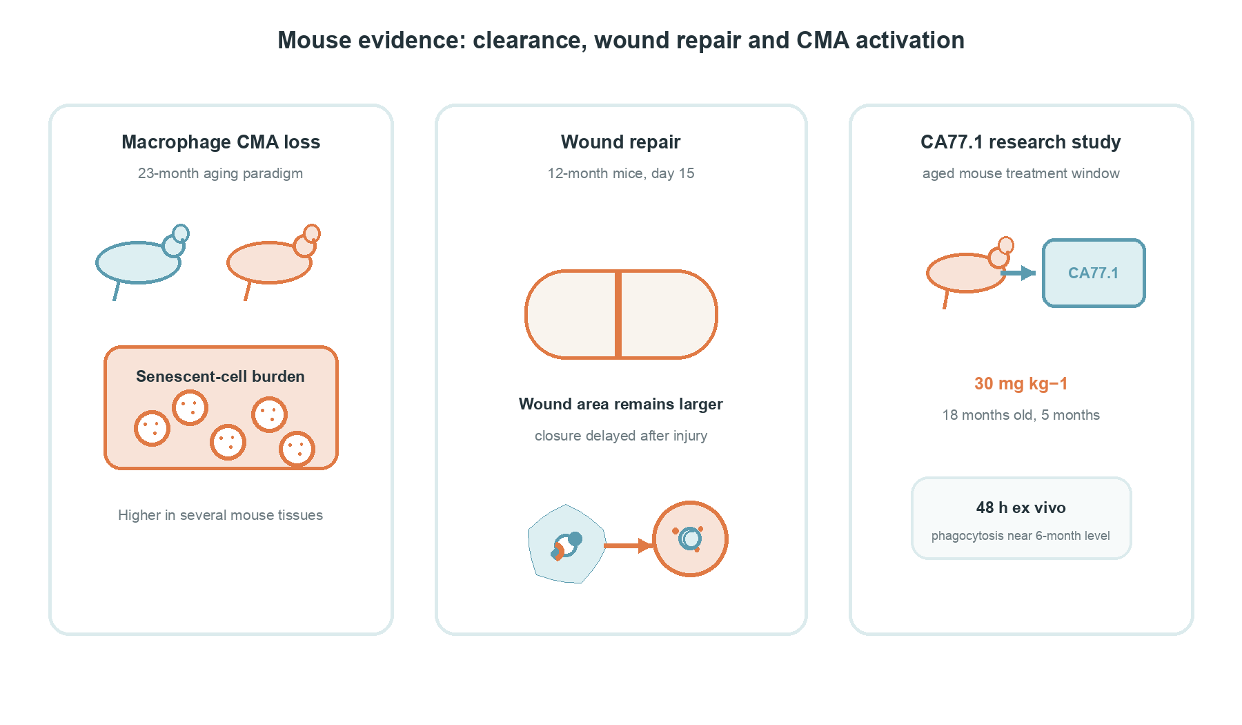
Macrophage-specific CMA deficiency elevates tissue senescent-cell burden and significantly delays wound healing in mice.
To evaluate in vivo consequences, the investigators deleted macrophage CMA in mice. These animals accumulated a substantially higher senescent-cell burden across multiple tissues during aging. In a wound-healing model using 12-month-old mice, wounds in macrophage CMA-deficient mice remained noticeably larger 15 days after injury, closure was delayed, and wound margins retained significantly higher numbers of p21-positive senescent cells.
Next, the authors administered oral CA77.1 at 30 mg kg−1 bodyweight daily to 18-month-old mice for 5 months. This chronic treatment reduced the accumulation of SA-β-gal-positive senescent cells in gonadal white adipose tissue, liver, and lung, alongside lowered transcription of canonical senescence markers. Furthermore, when bone-marrow-derived macrophages from 23-month-old mice were treated with CA77.1 for 48 hours ex vivo, their phagocytic capacity recovered to levels comparable to macrophages from 6-month-old mice. The study also reported attenuated disease severity in a mouse model of bleomycin-induced pulmonary fibrosis following pharmacological CMA activation.
Study scope: repairing CMA is not a proven human anti-aging therapy
The experimental evidence remains strictly confined to cell cultures and mouse models; this work is not a human clinical trial.
We must define the scientific boundaries clearly. Every data point in this report comes from cultured cells or laboratory mice. No human subjects participated, and no clinical efficacy or safety profile has been established. While pharmacological CMA stimulation represents an insightful mechanistic avenue, CA77.1 remains an experimental tool rather than an approved therapeutic compound or consumer anti-aging regimen.
Furthermore, senescent cells are not universally malicious. During acute tissue injury and wound healing, transient senescent cells provide vital paracrine cues that orchestrate cellular repair and tissue remodeling. Indiscriminately eradicating all senescent cells could compromise physiological regeneration. The ultimate goal of biomedical research is not to wipe out every senescent cell, but to understand why clearance systems fail and how to restore their natural balance.
Source: Sereda et al., Nature Aging (2026). https://doi.org/10.1038/s43587-026-01240-w