
In brief: Imagine a mature oocyte carrying roughly 100,000 mitochondria. It is not a quiet container. It is an energy factory that must support fertilization and the earliest stages of embryo development. When misfolded proteins accumulate inside those mitochondria, the first failure may be oocyte quality rather than the number of oocytes.
The short answer: the target is quality, not just quantity
This mouse study connects oocyte quality to mitochondrial protein cleanup and PGD2 signaling.
Published in Cell Death Discovery in 2026, the study first compared oocytes from young and reproductively old mice, then deleted Clpx specifically in oocytes with a Zp3-Cre system. With age, mouse oocytes had less ClpX and more unfolded proteins. The conditional knockout let the researchers separate a change that accompanies aging from a functional loss caused by removing ClpX.
ClpX is the unfoldase component of the mitochondrial ClpXP quality-control system. It unfolds damaged or misfolded proteins so that ClpP can process them. It is like a quality-control worker in a factory: without that first step, defective parts pile up and the energy system becomes harder to maintain.
The oocytes did not immediately disappear. They became less reliable under stress
The central result was not an immediate loss of oocytes in young knockout mice. It was a loss of competence across maturation, fertilization, and embryo development.
At two months, the Clpx-deficient mice did not show a clear difference in early oocyte or ovulated-oocyte counts. However, fertilization, germinal-vesicle breakdown, first polar-body extrusion, and later embryo development were reduced. Abnormal spindles, chromosome misalignment, and aneuploidy were more frequent. By 10 months, some follicle counts were also lower than in controls. The mitochondria showed higher reactive oxygen species, lower membrane potential, lower ATP, abnormal distribution, and fewer cristae.
You can think of this as a machine that still turns on but repeatedly stalls during a stress test. Counting oocytes alone misses the energy supply, chromosome organization, and cleanup capacity needed for development.
PGD2 translates mitochondrial stress into a trackable signal
The proposed chain begins with higher Ptgds and continues through PGD2 and two downstream signaling routes.
When the authors compared the Clpx dataset with other models of mitochondrial dysfunction, Ptgds was a shared upregulated transcript. Clpx-deficient oocytes also released more PGD2 and showed changes in DP1, PKA, cAMP, DP2, AKT, mTOR, and S6 signaling. Knocking down Ptgds improved first-polar-body extrusion and reduced spindle and chromosome abnormalities, supporting a functional role for the PGD2 axis rather than a purely incidental association.
The model is like an internal fault report moving from the mitochondrial workshop to the cell-signaling desk: protein handling fails first, PGD2 signaling rises next, and oocyte competence is affected afterward. However, the authors note that PTGDS/PGD2 may be a shared response to mitochondrial stress, not a ClpX-specific switch.
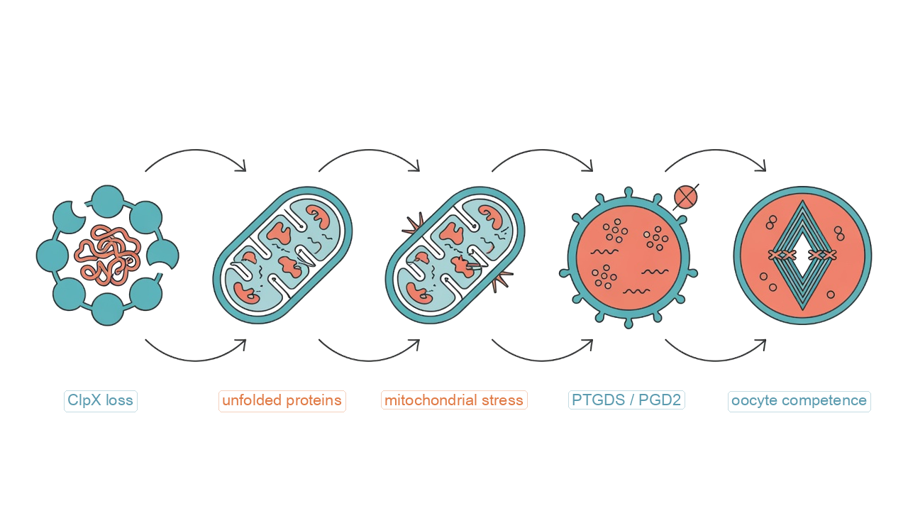
Figure 1. A study-grounded mechanism sketch. It shows the direction supported by the experiments and does not convert mouse findings into a proven human treatment.
AT-56 produced a signal, not a clinical fertility treatment
The PGD2-synthesis inhibitor AT-56 improved several maturation readouts in mouse experiments and in a small human oocyte in-vitro maturation experiment.
For mouse in-vitro maturation, the researchers used 100 μM AT-56. In vivo, they injected 10 mg/kg intraperitoneally into Clpx-deficient or reproductively old mice. Outcomes included partial improvements in germinal-vesicle breakdown, first-polar-body extrusion, fertilization, or embryo development. In human germinal-vesicle oocytes, 10 μM AT-56 was added for 24 hours, and maturation rates increased.
These are laboratory maturation outcomes, not proof that fertility treatment has succeeded. The human experiment did not report pregnancy, live birth, or postnatal health, and it was not large enough to assess safety. AT-56 should not be treated as an anti-aging drug or used as a supplement.
Scope and limitations set the boundary of the claim
The evidence boundary is straightforward: the main model was the mouse, while the human evidence stopped at oocyte maturation in vitro.
The authors list several limitations. ClpX and ClpP share the ClpXP complex, so their phenotypes may overlap. PTGDS/PGD2 may be a common mitochondrial-stress response. The efficacy, safety, and specificity of AT-56 still require validation. The human age comparison for CLPX expression included only 4 young and 3 older germinal-vesicle oocytes. That sample can support an initial signal, not a clinical-effect estimate.
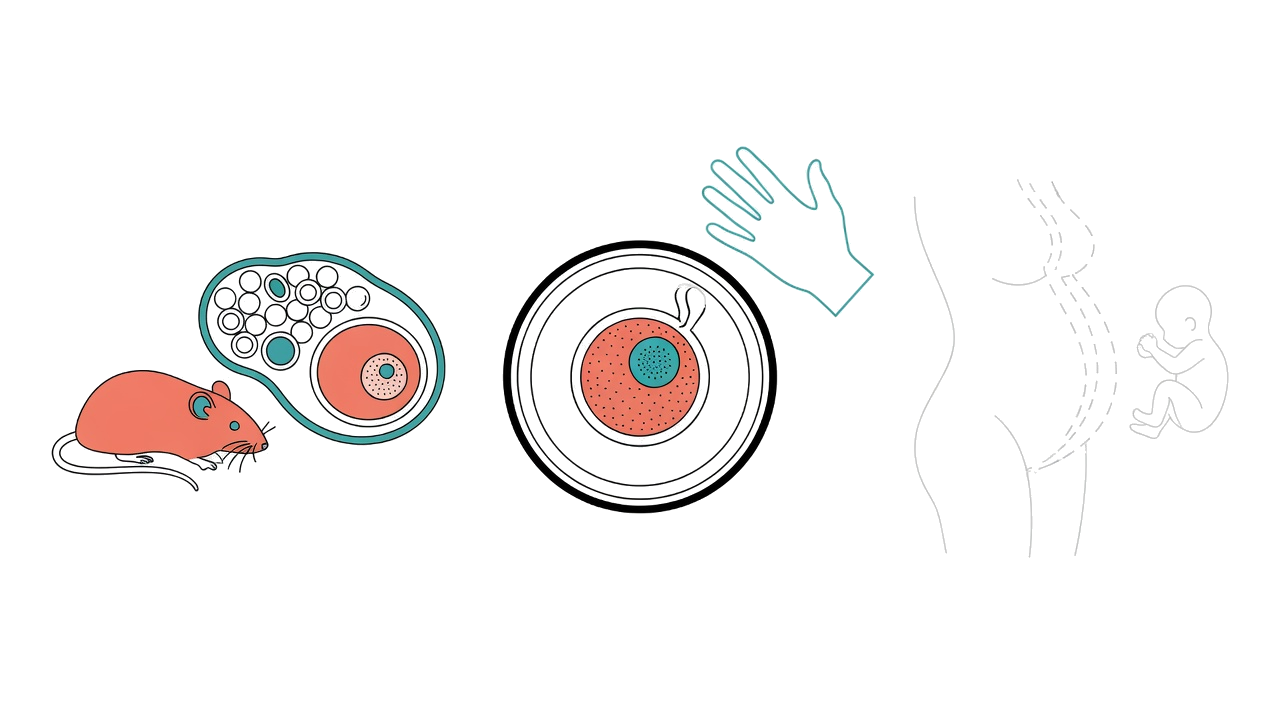
Figure 2. Pregnancy and live birth were not measured.
ClpX can be described as a cellular maintenance crew, but the study has not reached human treatment. Reading “better maturation” as “higher pregnancy or live-birth rates” would cross the evidence boundary. The most defensible value of this work is that it connects mitochondrial protein cleanup, PGD2 signaling, and oocyte quality in a way that can be tested further.
Source: Hua R, Hai Z, Zheng T, et al. Cell Death Discovery (2026). https://doi.org/10.1038/s41420-026-03335-0