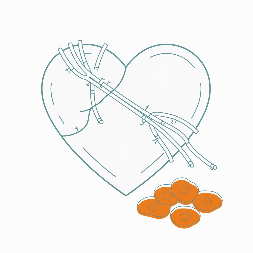
Imagine a second plumbing system sitting beside the heart's blood vessels. It is not an artery. It is more like a neighborhood storm drain: it clears extra fluid, proteins, and patrolling immune cells. Wagner and colleagues re-measured that network in aging hearts in Nature Cardiovascular Research (2026).
The heart has a drainage system of its own
Aging hearts are not only stiffer vessels or thicker muscle. Cardiac lymphatics run through the epicardium, myocardium, and endocardium, with the highest density at the epicardium. Capillaries use loose button junctions so fluid and cells can enter; collectors use tighter zipper junctions so the contents do not leak on the way out.
In mice, subendocardial lymphatics of the left ventricle thinned first, while right-ventricular vessels were relatively spared. Epicardial collectors widened. The pattern appeared in both sexes. Human spatial transcriptomics pointed in the same direction.
Buttons turn into zippers
When lymphatics thin, clearance slows and waste starts to pile up. Lymphatic density began to fall at 16 months. Macrophages rose by 18 months; T cells later. Macrophages in mediastinal lymph nodes dropped, consistent with impaired exit rather than failed production. Edema, fibrinogen, and Congo-red amyloid followed. Hypertrophy and fibrosis came later. Diastolic filling worsened; ejection fraction did not.
Correlation is not causation. Deleting VEGFR3 in lymphatic endothelium of young mice, or trapping VEGFC and VEGFD with soluble Flt4, cut lymphatic density by about 50%. Macrophages and fibrinogen rose, and diastolic function worsened, yet fibrosis and hypertrophy had not caught up after two months.
Nuclear IL-33 starts giving orders
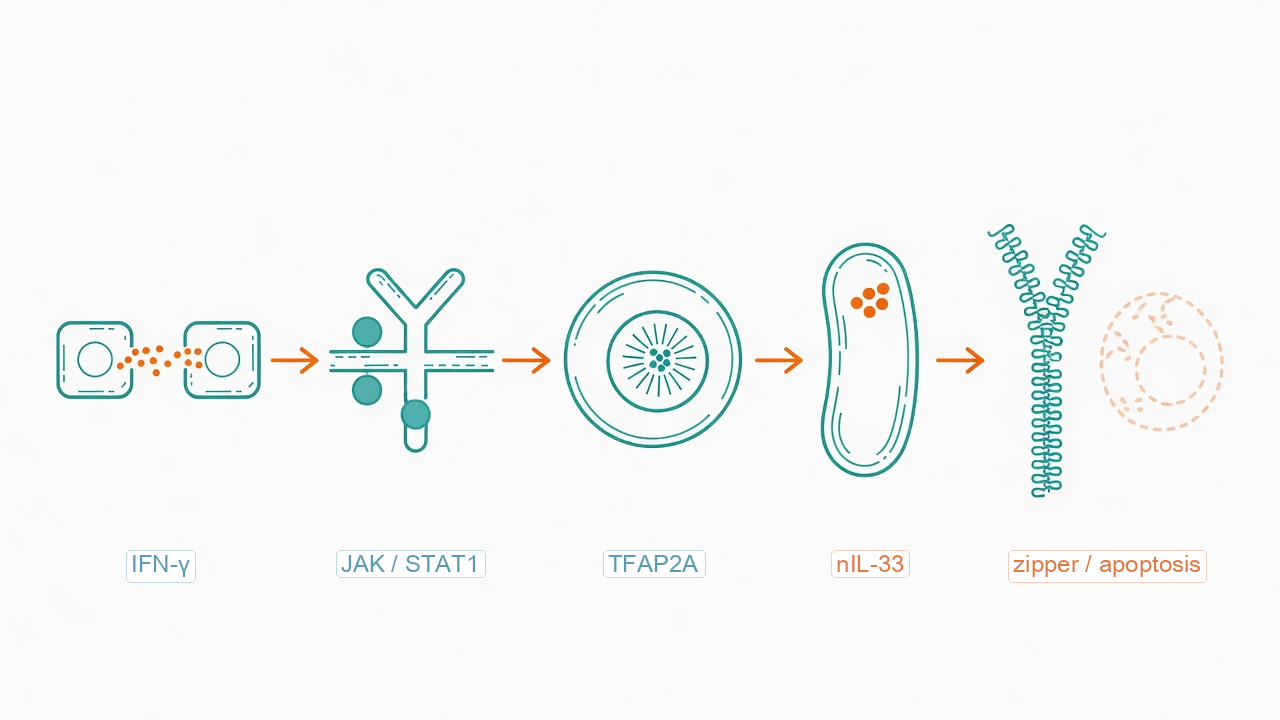
Aged lymphatic endothelial cells accumulate the nuclear form of IL-33. Single-cell RNA-seq found 49 genes up and 2 down in aged lymphatic endothelium. Il33 was a top hit, and the protein sat mainly in the nucleus. Secreted IL-33 can support lymphangiogenesis; nuclear IL-33 did the opposite in human dermal lymphatic endothelial cells, reducing proliferation, raising apoptosis, and increasing zipper junctions.
Among the cytokines tested, only IFN-gamma raised IL-33. T cells and NK cells in aged hearts expressed more Ifng, and T cells sat closer to lymphatics. JAK, STAT1, and TFAP2A sit on that path. Silencing Il33 with endothelium-targeted AAV9 in 18-month-old mice restored lymphatic density and reduced macrophages.
Restoring VEGFC did not rescue diastolic function
Overexpressing Vegfc in 18-month-old mice thickened lymphatics and reduced macrophages. Only VEGFC, among the growth factors tested, both stimulated proliferation and blunted IFN-gamma-driven IL-33. Cardiac Vegfc itself declined with age. After eight weeks of AAV9-Vegfc, nuclear IL-33 in lymphatics fell and PDPN-positive vessels rose. Diastolic function did not return.
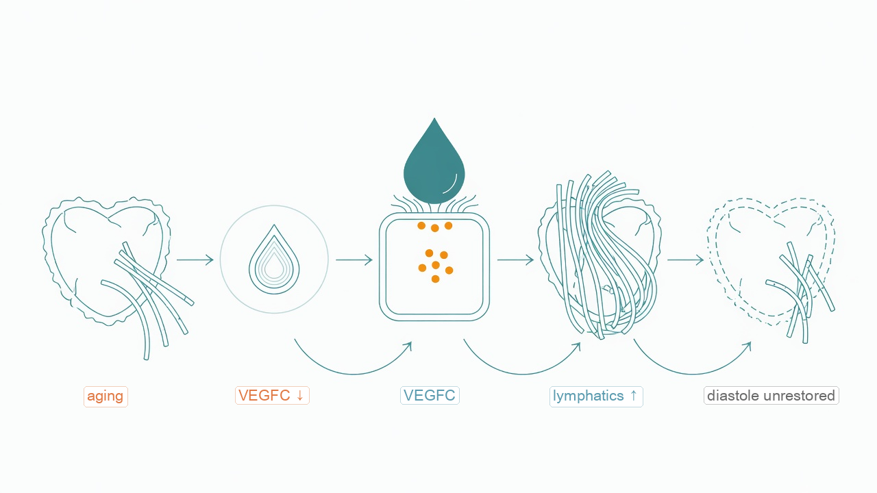
The study is a mouse and cell map, not a human trial. Sample sizes often ranged from 3 to 12 animals per group. Two months of lymphatic loss did not reproduce fibrosis. VEGFC rebuilt density without fixing the diastolic readout they measured. That is a route worth testing, not a reason to self-administer growth factors.