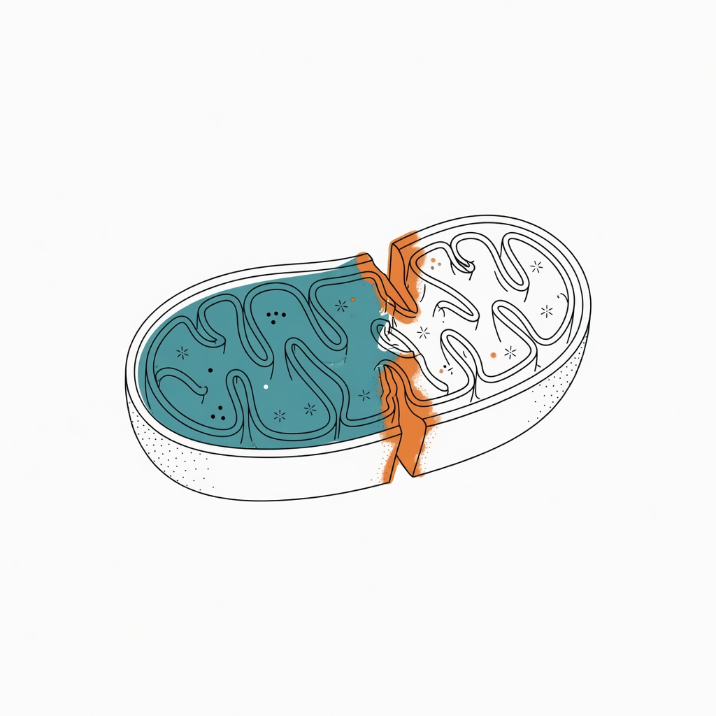
Imagine a power plant inside your cell. Mitochondria supply about 90% of ATP, yet they can also pile iron and oxygen radicals into a fire that eats membrane lipids. This is not science fiction. In 2012, Stockwell and colleagues named this death ferroptosis. Drug-resistant cancer cells can be especially afraid of that fire.
Ferroptosis is not a cousin of apoptosis
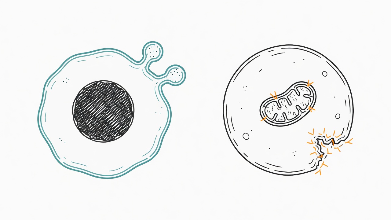
Ferroptosis is iron-dependent death by lipid peroxidation, and it does not look like apoptosis. Apoptotic cells show nuclear condensation and apoptotic bodies. Ferroptosis does not. Mitochondria shrink, cristae fade, and membranes rupture, more like metal losing structure after oxidation. Glutathione is used up, GPX4 stops working, and lipid peroxides accumulate. Once the membrane breaks, the cell cannot be rescued. Inducers fall into 2 classes. Class I agents such as erastin block system Xc- and starve cystine import. Class II agents such as RSL3 bind GPX4 directly. Both roads end with membranes being eaten.
Mitochondria are both power plant and tinder
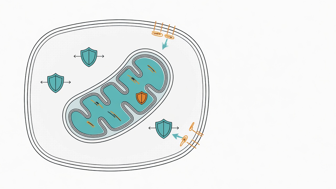
A 2025 review by Ding and colleagues puts mitochondria back at the center of ferroptosis control. Cells keep at least 4 firewalls: GSH-GPX4, FSP1-CoQ10, GCH1-BH4, and the intramitochondrial DHODH-CoQH2 system. The first three mostly repair peroxidized lipids in the cytosol or at membranes. DHODH reduces CoQ to CoQH2 inside mitochondria, like touching up paint inside the power plant. Mitochondria also control iron traffic. Iron enters through mitoferrin and is used to build heme and iron-sulfur clusters. Spare free iron can drive Fenton chemistry. ROS may then smash membrane lipids. CoQ is both an electron carrier and a lipophilic antioxidant. Extra CoQ can raise resistance to ferroptosis. Fusion-fission imbalance scrambles iron distribution. Mitophagy is like removing broken generators. Too slow, and tinder remains. Too fast, and iron may be released.
Why some therapy-resistant cancers are more sensitive
The review's working clue is that some cancer cells survive by shutting down ferroptosis, not only by resisting apoptosis. Mesenchymal, metabolically plastic cancer cells load more PUFA phospholipids into membranes, like stacking kindling on a wall. When GPX4 is pressed down, they become more fragile. p53 and BAP1 can also repress SLC7A11, cutting cystine supply and glutathione synthesis. But the same paper notes a counterexample: colorectal cancer cells with wild-type p53 can be less sensitive to erastin. The switch is not one-way. Under energy stress, AMPK issues orders, phosphorylates ACC, slows lipid synthesis, and lets cells briefly escape ferroptosis. Inhibiting the electron transport chain can also dampen the fire. Mitochondria do not only light the match, so we should not write them as a single switch. Some cells still stand after GPX4 fails because FSP1 or DHODH remains as backup.
How far is this from treatment
This paper is a literature review, not a human trial, and not a reason to self-medicate. The authors set the scope as mechanism mapping and translational imagination. Direct clinical evidence for targeting mitochondrial ferroptosis remains scarce. Inducing ferroptosis may injure neural or ischemic tissue, because the same path is linked to those diseases. No drug specifically hits mitochondrial GPX4. DHODH inhibitors mostly remain in cells and mouse xenografts. The molecular steps are still incomplete, and the toxicity boundary is unclear. This is not a verdict. It is a map waiting to be tested, reminding us that resistance is not only about apoptosis. The answer is still on the road.