Mechanical ventilation is one of modern medicine's most important life-saving tools. For patients in respiratory failure, it is the bridge between death and survival. But that bridge may carry unexpected traffic in the opposite direction.
A 2026 paper in the Journal of Advanced Research asks a discomforting question: could the mechanical stretch imposed by ventilators on alveolar cells be contributing to the very pulmonary fibrosis it's being used around?
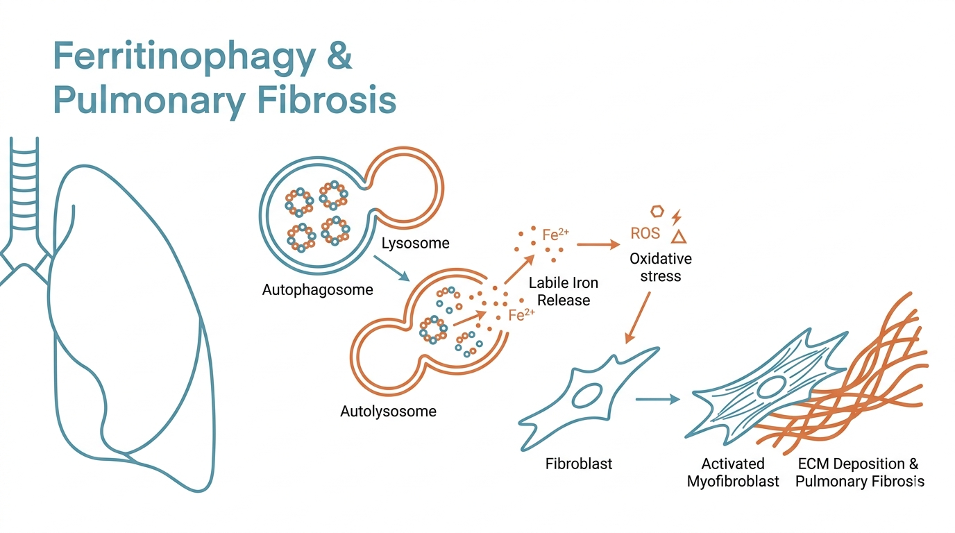
Takeaway: Huang's team found that mechanical ventilation — at least in high-tidal-volume models — can trigger NCOA4-mediated ferritinophagy in alveolar epithelial cells. This releases iron from ferritin stores, promotes lipid peroxidation, and drives ferroptosis. Dying cells then release ferritin-containing extracellular vesicles (EVs) that are taken up by fibroblasts, activating them and promoting extracellular matrix deposition. Inhibiting ferritinophagy reduced both ferroptosis and fibrosis in these models.
The Unexpected Culprit: Iron Dysregulation
Pulmonary fibrosis is often described as abnormal wound healing — the lung scars where it should repair. But the cellular events that initiate and propagate that scarring process are still being defined.
This paper introduces a pathway that hasn't received much attention in the fibrosis field: iron metabolism. The research team used two models — a high-tidal-volume mouse ventilation model and mechanically stretched alveolar epithelial cells in culture. In both systems, mechanical stress disrupted iron homeostasis in ways that cascaded into fibrosis.
Single-cell analysis revealed that genes associated with ferroptosis suppression — including Gpx4 (glutathione peroxidase 4) and Fth (ferritin heavy chain) — were downregulated in mechanically stressed alveolar cells. These are part of the cell's natural defense against iron-driven oxidative damage. When they fall, the defensive perimeter gets thinner.
The Three-Step Mechanism
The paper proposes a sequential pathway that converts mechanical force into fibrotic tissue remodeling.
Step 1: NCOA4-Mediated Ferritinophagy
Ferritin is the cell's iron storage protein — it sequesters free iron to prevent it from participating in damaging chemistry. NCOA4 is a cargo receptor in the autophagy pathway that selectively delivers ferritin to autolysosomes for degradation. Under normal conditions, this process is regulated and serves iron recycling.
In mechanically stressed cells, this system becomes dysregulated. The paper shows that the ANG II/AGTR1 signaling axis activates NCOA4, and that NCOA4 itself promotes ferritin phase separation — essentially clustering ferritin molecules together, making them more efficiently targeted for autophagic degradation. The iron vault gets dismantled.
Step 2: Ferroptosis
When ferritin is degraded at excessive rates, free iron is released into the cytoplasm. Iron in its labile, reactive form drives the Fenton reaction and lipid peroxidation. When lipid peroxidation overwhelms the cell's antioxidant defenses — particularly GPX4 activity — the cell undergoes ferroptosis, a regulated form of cell death characterized by oxidative lipid damage.
Unlike apoptosis, ferroptosis doesn't produce neat cell fragments. It releases cellular contents — including proteins and organelles — into the local environment. And this is where the story becomes particularly concerning.
Step 3: Ferritin-Carrying EVs
As alveolar epithelial cells undergo ferroptosis, they release extracellular vesicles containing ferritin. These vesicles are taken up by nearby fibroblasts. Once inside the fibroblast, the ferritin-loaded EVs deliver iron — effectively extending the iron dysregulation from dying epithelial cells to living fibroblasts.
Activated fibroblasts are the central effectors of pulmonary fibrosis: they produce collagen and extracellular matrix components that replace normal lung architecture with stiff, non-compliant scar tissue. By showing that ferritin-containing EVs can activate fibroblasts, the paper describes a mechanism by which the injury doesn't stay localized — it propagates spatially via the EV cargo delivery system.
Evidence That the Pathway Is Real
The mechanistic story is coherent, but the evidence supporting it is also multi-layered.
The team demonstrated that inhibiting ferritinophagy — either pharmacologically with chloroquine or genetically using AAV-mediated NCOA4 knockdown — reduced ferroptosis markers, lowered EV ferritin content, and attenuated fibrosis in the mouse model. This reverse-validation confirms the pathway's functional importance rather than just its correlation.
Importantly, they also showed that ferritin phase separation — the biophysical clustering step that makes NCOA4-mediated targeting more efficient — was mechanistically upstream of the downstream iron release and ferroptosis. This provides a specific molecular target that hadn't been implicated in ventilator-induced lung injury before.
Limitations and Clinical Distance
This paper is mechanistically rich but clinically distant. Several caveats are essential.
First, the high-tidal-volume ventilation model is deliberately injurious — it is not the same as lung-protective ventilation strategies (lower tidal volumes, appropriate PEEP) that are now standard in most ICU settings. The model is useful for interrogating mechanisms under stress conditions but doesn't directly predict outcomes in contemporary clinical practice.
Second, real patients on ventilators are a heterogeneous population with different underlying conditions, different ventilator parameters, and different systemic inflammatory states. The clean mechanistic story in two model systems will encounter substantial biological noise in the clinic.
Third, the interventions used to confirm the pathway — chloroquine, AAV knockdown — are proof-of-concept tools, not clinical therapies. Whether any of this translates to actionable pharmacological intervention requires substantially more development.
Finally, whether ferritin-containing EVs in the circulation or bronchoalveolar fluid could serve as clinical biomarkers for fibrosis risk in ventilated patients remains entirely unexplored.
The Contribution: A Molecular Map
Despite these limitations, the paper makes a genuine contribution. It takes the observation that mechanical ventilation can worsen pulmonary fibrosis — a clinical reality in ventilator-associated lung injury — and provides a specific, testable molecular explanation: NCOA4 ferritinophagy → free iron release → ferroptosis → ferritin-EV-mediated fibroblast activation.
This is a map, not a treatment. But maps are what allow therapeutic development to proceed with direction rather than intuition. The iron metabolism angle in pulmonary fibrosis is underexplored, and this paper provides solid ground for future investigation.
For clinicians: this reinforces the importance of lung-protective ventilation strategies. For researchers: ferritinophagy, ferroptosis, and EV-mediated iron transfer are worth examining in other fibrotic diseases where mechanical or oxidative stress is a driver.
References
- Huang et al. (2026). NCOA4-Mediated Ferritinophagy Induces Ferroptosis and Enriches Ferritin-Containing EVs via Ferritin Phase Separation to Promote Mechanical Ventilation-Induced Pulmonary Fibrosis. Journal of Advanced Research. doi: 10.1016/j.jare.2025.07.043