TL;DR: Iron is essential. But under the wrong conditions, it ignites a chain reaction inside your cells that looks, at the molecular level, exactly like rust. This process — called ferroptosis — was only named in 2012. Scientists are now finding it may be one of the core engines of aging.
You thought iron was just a nutrient
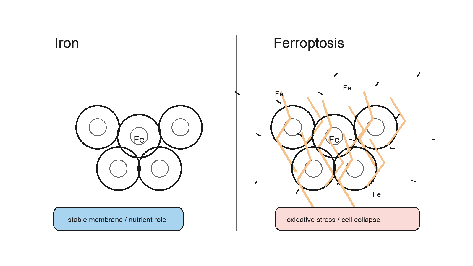 Figure 1: Iron is essential for life, but in excess or the wrong context it becomes a trigger for oxidative cell death.
You take iron supplements. You eat spinach. You worry about anemia. What you probably do not know is that the same element making your blood red holds a second key — one that, under specific conditions, lights a slow, silent fire inside your cells.
That fire has a name: ferroptosis (iron death).
Not a heart attack. Not a tumor. Your cells dissolving from the inside, driven by a collision between excess iron and oxidizable fat. Scientist Brent Stockwell named this death mechanism in 2012. In less than fifteen years, it has moved from a biochemistry curiosity to a central topic in aging research.
Are your legs getting weaker? Your joints stiffer? You may never have heard this word — but the process may already be happening in your tissues.
A chemical fire inside the cell membrane
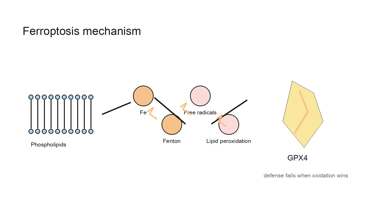 Figure 2: GPX4 is one of the cell's key defenses against runaway lipid peroxidation and ferroptosis.
Picture your cell membrane as a wall — a bilayer of phospholipid molecules, each one flexible because of its unsaturated fatty acid tails. That flexibility is a liability.
Unsaturated fatty acids oxidize easily. When free iron accumulates inside a cell, it drives the Fenton reaction: iron converts ambient oxygen into highly reactive free radicals. Those radicals ignite the fatty acid chains in the membrane, triggering a spreading chain reaction called lipid peroxidation.
The wall starts to crack.
Cells have a defense: GPX4 (glutathione peroxidase 4), an enzyme that acts as the cell's fire suppressor, neutralizing lipid peroxides before they spread. When GPX4 activity declines — or when iron overload overwhelms it — ferroptosis follows.
A 2025 landmark review in Physiological Reviews (Zheng et al.) frames ferroptosis not as a binary switch but as a dynamic equilibrium between iron metabolism, lipid metabolism, and antioxidant defense. Disrupt any one of the three, and the fire starts.
Aging pours fuel on the fire
Three things happen as your body ages — and all three feed ferroptosis.
Senescent cells learn to escape death.
Senescent cells are the "zombie cells" of your tissues: they stop dividing but refuse to die. A 2025 study in Nature Communications (Zhou et al.) found that senescent cells upregulate a protein called IFI16, which in turn promotes HMOX1 expression, making these cells resistant to ferroptosis. The zombies do not just survive — they develop a shield against the very fire meant to clear them. Meanwhile, they keep secreting pro-inflammatory signals, loading their neighbors with oxidative stress.
Aged macrophages ignite muscle ferroptosis.
A 2025 Nature Aging study (Xiang et al.) placed senescent macrophages into skeletal muscle microenvironments. The result was clear: senescent macrophages trigger ferroptosis in muscle cells, driving sarcopenia (muscle loss), which then cascades into osteoarthritis. The "natural" leg weakness of aging may have a cellular arsonist behind it.
Mitochondria shift fuel strategy — and make cells more fragile.
Research published in Science Advances in 2024 (Yamauchi et al.) showed that aging cells increasingly rely on mitochondrial fatty acid oxidation for energy. This metabolic shift reinforces the senescence phenotype — and it floods the cellular environment with the very lipid substrates that ferroptosis requires.
This is not coincidence. It is a mechanism: at the molecular level, aging is, in part, a slow process of rusting from within.
Can blocking ferroptosis extend healthy life?
Researchers have tested this across multiple species.
A 2024 Advanced Science study (Fu et al.) ran ferroptosis-inhibition experiments in nematodes, fruit flies, and mice. The results were consistent across all three: suppressing ferroptosis not only extended lifespan but improved aging phenotypes across the board — mobility, tissue integrity, resistance to oxidative stress. Nematode lifespan increased by approximately 25%. Skeletal muscle atrophy was meaningfully reduced in aged mice.
What about humans?
Clinical translation is early-stage. But the research landscape points toward several practical directions: supplementing CoQ10 and Vitamin E to reinforce the membrane's antioxidant barrier; N-acetylcysteine (NAC) as a precursor substrate for GPX4 activity; and managing iron load — particularly avoiding excess non-heme iron supplementation — as a potential modifiable risk factor.
This is not a case against iron. It is a reminder that iron metabolism is more precise than most people appreciate.
Rust can be slowed
Aging is not a moment. It is an accumulation. Ferroptosis does not arrive all at once — it advances one oxidative insult at a time, one chain reaction at a time.
Every serving of dark leafy vegetables, every bout of aerobic exercise giving your mitochondria a cleaner energy source, every reduction in chronic inflammation — these are interventions at the cellular level, whether or not they feel like it.
Scientists are working toward precise pharmacological targets: GPX4 stabilizers, ACSL4 inhibitors, clinical applications of Ferrostatin-1. Until that era arrives, the lifestyle levers available to you are more mechanistically grounded than they may appear.
Rust is not destiny. It is a process. Processes can be slowed.
References
- Zheng, J. et al. (2025). Ferroptosis: when metabolism meets cell death. Physiological Reviews. https://doi.org/10.1152/physrev.00031.2024
- Fu, Y. et al. (2024). Inhibition of ferroptosis delays aging across species. Advanced Science. https://doi.org/10.1002/advs.202416559
- Xiang, X. et al. (2025). Senescent macrophages induce ferroptosis in skeletal muscle cells. Nature Aging. https://doi.org/10.1038/s43587-025-00907-0
- Zhou, H. et al. (2025). IFI16 promotes HMOX1-dependent evasion of ferroptosis in senescent cells. Nature Communications. https://doi.org/10.1038/s41467-025-56456-y
- Yamauchi, S. et al. (2024). Mitochondrial fatty acid oxidation drives cellular senescence. Science Advances. https://doi.org/10.1126/sciadv.ado5887