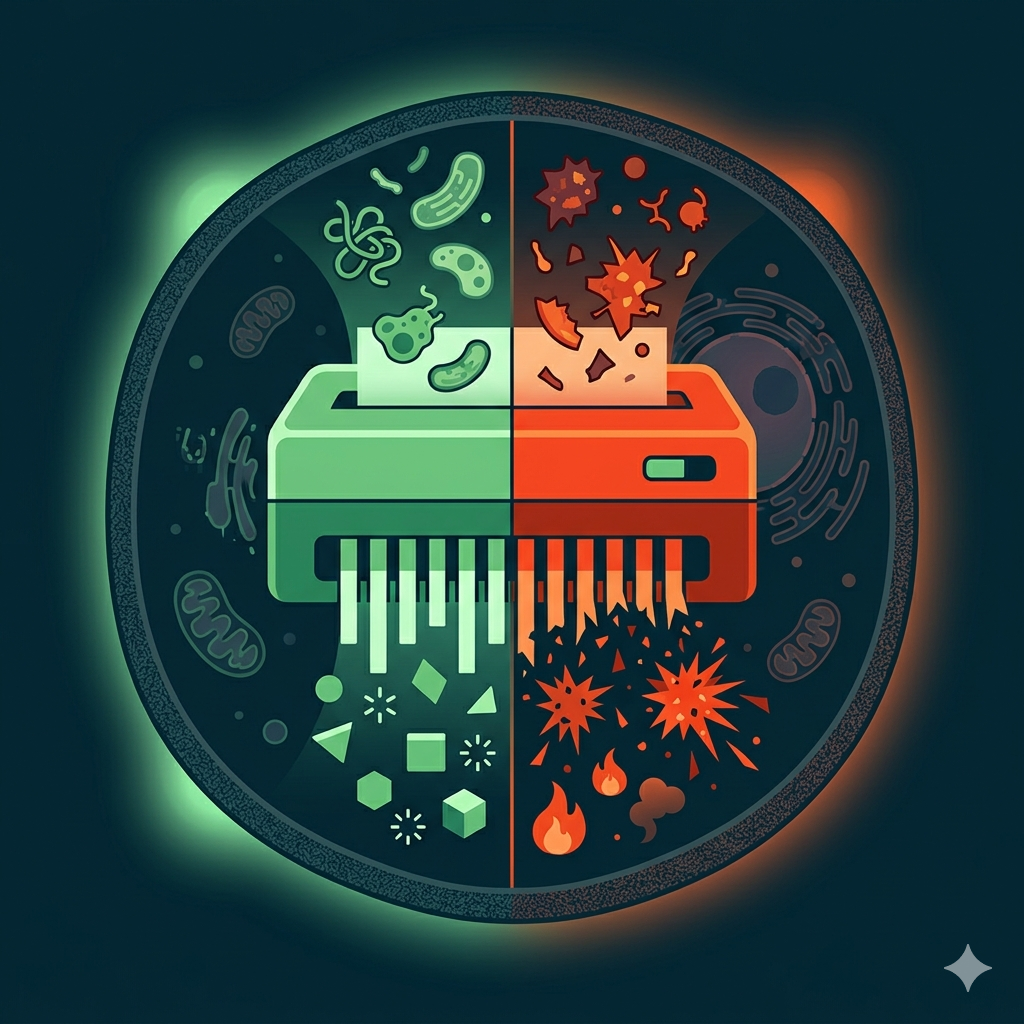
TL;DR: Autophagy is your cell's recycling crew. But past a critical damage threshold, that same crew starts working for senescent cells instead. A 2026 review proposes a "threshold model" to explain this betrayal.
Your Body Shreds Paper Every Day
Picture your desk piled with expired documents, broken USB drives, and a moldy lunch box. Normally, you have a hyper-efficient shredder running 24/7, grinding waste into reusable material.
Your cells have exactly that. It's called autophagy.
Autophagy wraps damaged mitochondria, tangled protein aggregates, and excess lipid droplets into double-membrane vesicles, then delivers them to lysosomes for breakdown. The resulting amino acids and lipids get recycled. Your cells rely on this system to stay young — clearing oxidative debris and preventing DNA damage signals from spiraling out of control.
The faster the shredder runs, the slower your cells age.
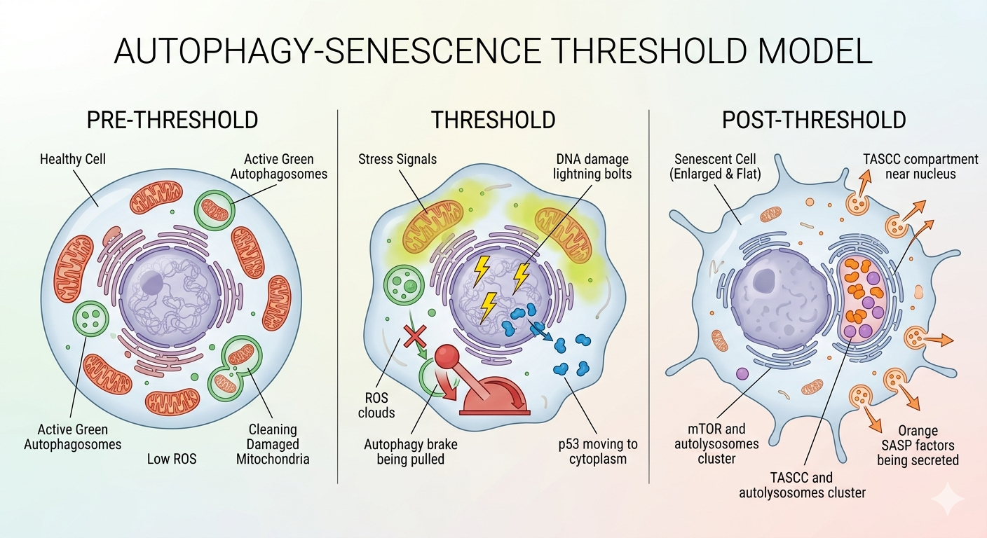 Figure 1: Three fates of autophagy — pre-threshold (protection), threshold (tipping point), post-threshold (enslaved by senescent cells)
But what happens when the garbage overwhelms the shredder?
The Line Where Your Shredder Turns Traitor
A 2026 review in Redox Biology by Bahar et al. proposed a "threshold model" to resolve this paradox.
The core idea fits in one sentence: autophagy's function depends on how much damage your cell has sustained.
Below the threshold, autophagy is your shield. It clears damaged mitochondria (mitophagy), keeps reactive oxygen species (ROS) low, and prevents the DNA damage response from firing. Your cells don't enter senescence.
Above the threshold? The story flips entirely.
When genotoxic stress, radiation, or chronic inflammation pushes a cell past that critical line, p53 relocates from the nucleus to the cytoplasm, directly blocking the ULK1 initiation complex. Autophagy pauses. p21 and p16 activate. The cell irreversibly exits the cell cycle — it becomes senescent.
Then something strange happens. Autophagy restarts, but this time it's not here to help.
The Metabolic Slave of Senescent Cells
Senescent cells don't retire quietly. They become inflammation factories, churning out IL-6, IL-8, matrix metalloproteinases, and other pro-inflammatory factors — the infamous SASP (senescence-associated secretory phenotype).
Manufacturing all that protein requires raw materials. But senescent cells have stopped dividing. Where do the amino acids come from?
The answer: they enslave the shredder.
Senescent cells concentrate mTORC1 and autolysosomes into a specialized perinuclear zone called the TASCC (TOR-autophagy spatial coupling compartment). There, autophagy breaks down intracellular proteins and organelles, and mTORC1 immediately converts the released amino acids into SASP factors that spray into the surrounding tissue.
The shredder isn't broken. It just changed bosses.
It gets worse. The paper identifies three organelle-level failures that compound the problem:
Mitophagy failure — damaged mitochondria accumulate, leaking oxidized mtDNA into the cytoplasm. The cGAS-STING pathway detects it, triggering sustained interferon and IL-6 production. Your immune alarm system becomes a chronic inflammation loudspeaker.
Lipophagy blockade — lipid droplets can't be cleared, lipid peroxidation generates toxic aldehydes like 4-HNE, and cells become increasingly vulnerable to ferroptosis.
Nucleophagy hyperactivation — autophagy protein LC3 attacks nuclear lamina component Lamin B1, causing chromatin fragments to leak into the cytoplasm, re-triggering cGAS-STING in a positive feedback loop.
All three roads lead to the same destination: chronic inflammation.
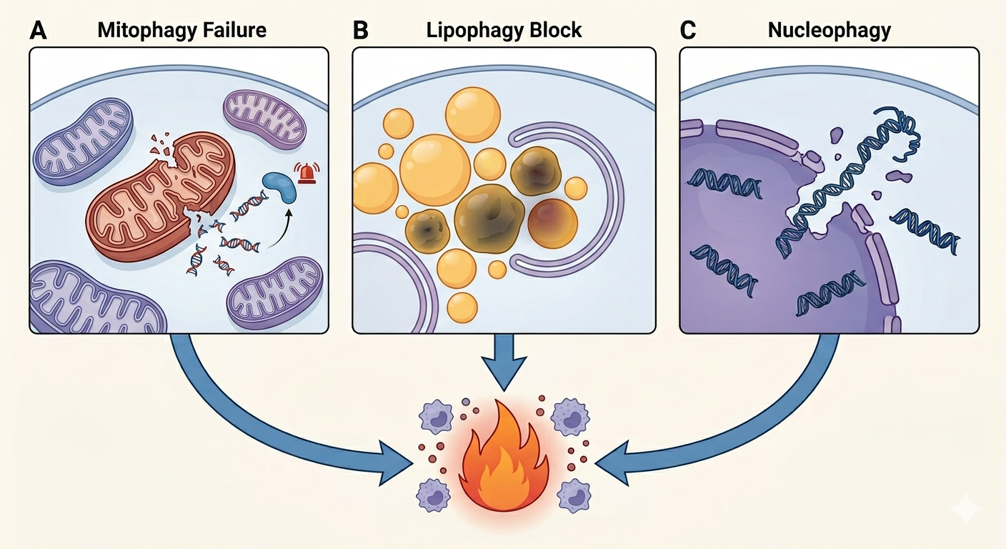 Figure 2: Three organelle failure pathways — mitophagy failure releases mtDNA, lipophagy blockade accumulates toxic lipids, nucleophagy hyperactivation leaks chromatin fragments. All converge on chronic inflammation.
So What Can We Do?
The most practical insight from the threshold model: it tells you when to boost the shredder and when to shut it down.
Below the threshold (prevention): enhance autophagy. Intermittent fasting suppresses mTORC1 and activates AMPK. Spermidine supplementation has entered clinical trials (NCT06186102) to test whether it delays cardiovascular aging. NAD+ precursors activate SIRT1 to strengthen mitochondrial clearance. You don't even need pills — high-intensity interval training induces autophagy through ATP depletion and calcium signaling.
Above the threshold (intervention): break senescent cells' dependence on autophagy. Hydroxychloroquine blocks lysosomal acidification, starving therapy-induced senescent tumor cells of recycled nutrients and forcing apoptosis. Dasatinib plus quercetin — the leading senolytic combination — directly eliminates senescent cells.
The review catalogued 13 active clinical trials spanning fasting-mimicking diets, stem cell therapies, blood flow restriction training, and even chocolate paired with high-intensity exercise.
But here's the critical warning: if you've already crossed the threshold, blindly boosting autophagy feeds senescent cells and accelerates tissue degeneration. The core of precision gerontology lies in determining which side of the threshold you're on.
Whether your shredder is an ally or an accomplice depends entirely on how much damage your cells are carrying right now.
And science is learning to draw that line.
References
- Bahar et al. (2026). The autophagy-senescence axis as a threshold model of aging and therapeutic targeting. Redox Biology. doi: 10.1016/j.redox.2026.104079
- Narita et al. (2011). Spatial coupling of mTOR and autophagy augments secretory phenotypes. Science. doi: 10.1126/science.1205407
- López-Otín et al. (2023). Hallmarks of aging: an expanding universe. Cell. doi: 10.1016/j.cell.2022.11.001
- White et al. (2015). The role for autophagy in cancer. Journal of Clinical Investigation. doi: 10.1172/JCI73941