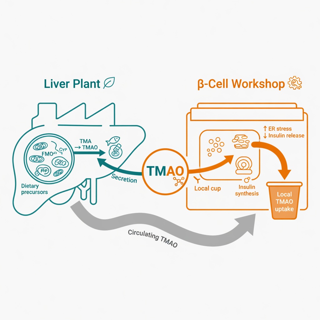
Why does the same TMAO wear two reputations?
If you only follow blood reports, TMAO (trimethylamine N-oxide) often plays the villain: gut microbes turn choline-like inputs into TMA, hepatic FMO3 oxidizes it to TMAO, and higher circulating levels line up with stacks of cardiometabolic risk papers.
Your body is not one factory.
Pancreatic β-cells can make TMAO through FMO3 as an autocrine/intracellular metabolite that restrains NF-κB-linked senescence and inflammation and helps keep glucose-stimulated insulin secretion (GSIS) online. In 2026, Wang, Yang and colleagues mapped this “shop-floor workshop” in Nature Communications: same molecule, different address from the river in your bloodstream.
A high river level is not the same story as the water left in the cup on the bench.
β-cells run their own FMO3 workshop
Textbooks paint FMO3 on the liver. The authors chased stable isotopes: feed d9-TMA, watch who builds d9-TMAO. In middle-aged male human samples, islet explants could synthesize TMAO faster than liver explants; islets from people with type 2 diabetes ran this line more weakly, while hepatic capacity looked similar between non-diabetic and diabetic donors.
Primary mouse cells match the “equipped, but not liver-clone” picture: islet Fmo3 mRNA sat about 99% lower than hepatocytes from the same animal, yet protein still reached 50–60% of male hepatocyte levels, without the strong sexual dimorphism classic for liver FMO3. Wipe FMO3 out only in β-cells and intracellular TMAO falls while circulating and hepatic TMAO need not crash: cup water, not a river gauge.
That sits beside an older hint: antisense knockdown that cuts hepatic FMO3 by roughly 90% often drops circulating TMAO by only about 20–30%. The liver is large. It is not alone.
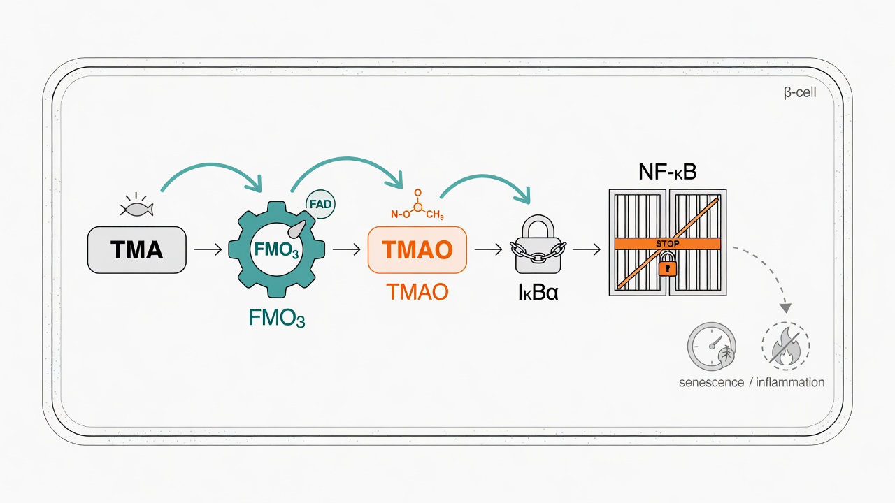
The β-cell FMO3–TMAO axis stabilizes IκBα, the latch on the NF-κB door, so less of the senescence/inflammation program is transcribed. For anyone whose meals depend on timed insulin pulses, that is on-site maintenance, not a press release from the central plant.
From glucose intolerance at week 12 to the IκBα latch
At eight weeks, β-cell FMO3 knockout mice still looked largely normal for glucose tolerance, insulin secretion, and insulin sensitivity. Mild glucose intolerance crept in around week 12, driven mainly by weaker GSIS, not a sudden surge of peripheral insulin resistance. Isolated islets at week 16 still showed the GSIS defect. Silencing Fmo3 in INS-1E cells broke GSIS too; adding TMAO back lifted total insulin content to about 50% of scramble controls; half a repair, not a full reset.
The ageing slice is louder. RNA-seq on islets from 48-week-old knockouts found 1353 differentially expressed genes (990 up, 363 down; |log2 fold change| ≥ 1, adjusted p ≤ 0.05). NF-κB-linked cytokine transcripts rose; among canonical senescence markers, p53 and p21 (Cdkn1a) moved up while p16 (Cdkn2a) did not follow the same script. β-galactosidase signal, p21 protein, and phospho-RelA activity brightened inside islets. TMAO add-back nudged those arms back.
The latch story is direct: TMAO binds IκBα, slows its disposal, limits NF-κB nuclear entry, and trims the transcriptional fuel for senescence and inflammation. PERK, a frequent character in hepatic TMAO tales, did not star in this β-cell cast.
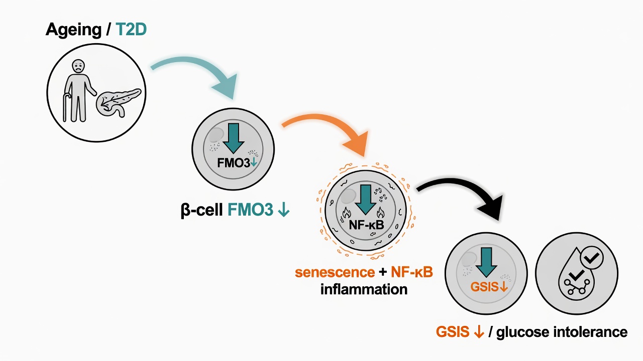
Human tissue is present: restoring FMO3 in aged human islets lowered NF-κB activation and senescence marks. Weaker TMAO synthesis in diabetic islets reads like the workshop losing power first.
You have also heard the opposite chorus: higher circulating TMAO, choline diets that push blood TMAO up, hepatic FMO3 making metabolic trouble, even reports that TMAO harms β-cell calcium signaling through inflammatory routes. Those studies mostly sample the river or systemic add-ons. This paper samples the cup the β-cell fills for itself. Same formula; different address and dose language; conclusions that look like a quarrel until you label the map.
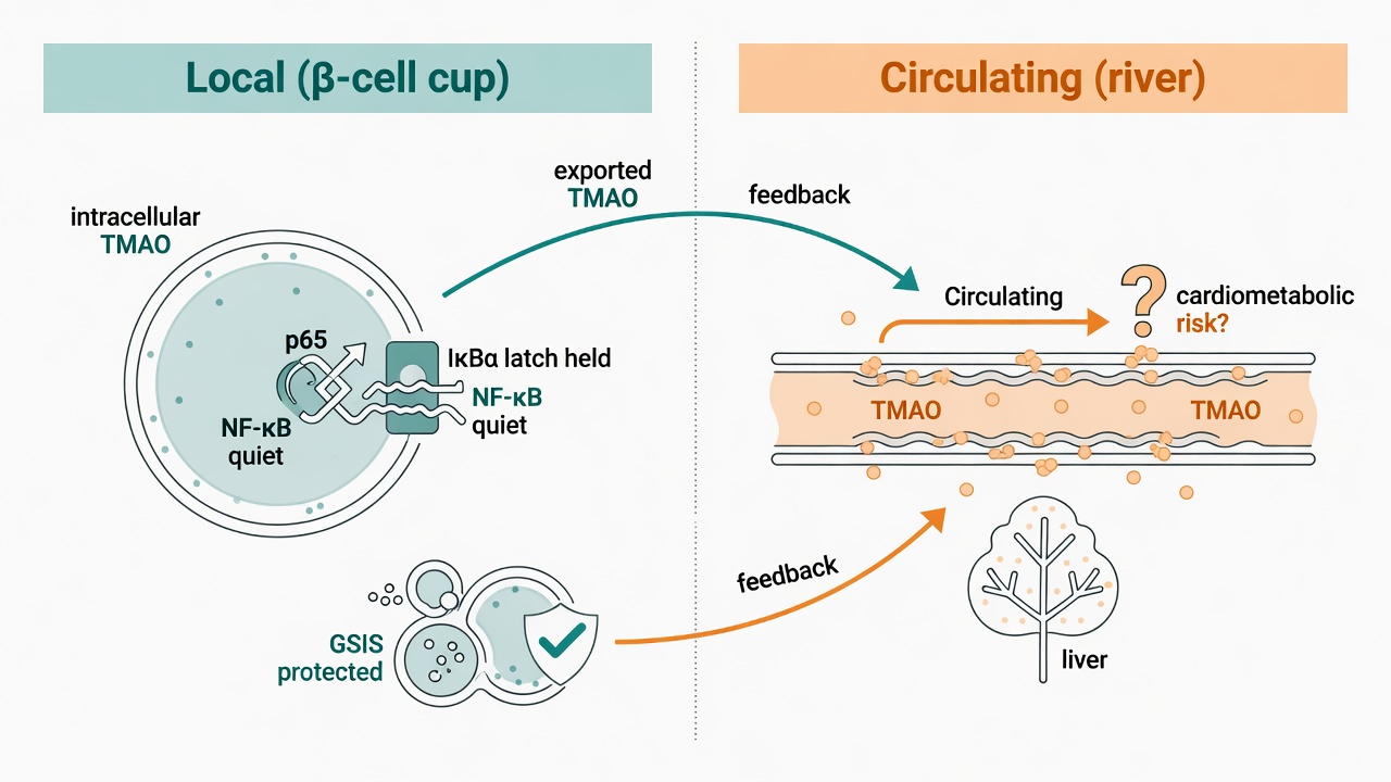
Scope stays tight: mouse conditional genetics, cell lines, human islet explants and aged-islet rescue, not a license to swallow TMAO powder. The human isotope arm leaned on middle-aged men; RNA-seq and tolerance time points are not one calendar day. Translating “week-12 mouse intolerance” into “middle-age human FMO3 failure” tears the scale bar off the map.
The next step is not flooding the bloodstream
If the axis holds, the more honest drug fantasy is repairing the β-cell’s own FMO3 line or carefully tuning local TMAO vocabulary in the islet niche, not jacking circulating TMAO as a longevity tonic. A single high blood TMAO value is not a certificate that your β-cells are thriving.
Your islets must meter insulin across every glucose swing. If the shop-floor workshop browns out in ageing and diabetes, prettier numbers from the central plant will not fix the post-meal hour. When you read TMAO papers, ask first: river, or cup?